Eye-care professionals
A CLINICAL INVESTIGATION DESK
Ortho-K Fitting: A Structured Assessment and Troubleshooting Guide
For clinicians: investigate Ortho-K fitting concerns through safety checks, reliable measurements and the exact design’s instructions before adjustments.Follow the safety-first investigation route ↓
Assess ocular health, refraction, corneal shape and the family’s ability to manage overnight lenses before selecting a design. Troubleshoot symptoms, lens condition and repeatable measurements before changing parameters. Follow the exact manufacturer’s fitting guide and clinical findings; a generic pattern diagram cannot prescribe a lens change.
On this page
Decide whether this is a safety problem before a fitting problem
An Ortho-K review can begin with blur, a map that looks different, discomfort or a lens that is hard to remove. Do not collapse those presentations into the same parameter-change question. First establish what the patient is experiencing and examine the eye appropriately.
Symptoms such as pain, redness, light sensitivity or a change in vision need prompt clinical assessment. A suspected adverse event is not an opportunity to keep the lens on while trying another fitting adjustment. Follow the relevant urgent-care pathway and product instructions. [2]
For a comfortable patient with a performance concern, record the timing and task: blur on waking, variable afternoon vision, glare, or a change limited to one eye. Note wear history, removal timing, current correction and any recent change in health, supplies or routine.
A useful troubleshooting note separates the observation, the checks performed and the decision. “Poor fit” is a conclusion. A repeatable finding, documented with its context, gives the next clinician something to assess.
Establish a reliable baseline
Assess suitability before selecting a lens design. The examination should address the clinical question, ocular health, refractive findings, corneal characteristics, visual needs and the practical ability to deliver care. Discuss alternatives and the role of the caregiver.
Record the instrument, method and quality of baseline measurements. Corneal maps should be reproducible enough to support the decisions being made. Save the original data and relevant settings so later comparisons do not depend on a screenshot without a scale or acquisition context.
The IMI instrumentation report distinguishes the information available from corneal-shape measurement, refraction and ocular biometry. These tests answer related but different questions. A topographic change does not itself measure axial growth, and an eye-length measurement does not describe lens centration. [3]
Ask about previous lens wear and obtain appropriate records. When a baseline is affected by prior treatment or another factor, document the limitation. Do not present an uncertain baseline as though it were a clean pretreatment comparison.
VISUAL GUIDE 1 / 3
Symptoms first, then the fitting investigation
Follow the clinical urgent-care pathway and relevant lens instructions.
Check timing, vision, eye health, lens history and measurement quality.
Use the instructions for the exact design
“Ortho-K lens” describes a category, not a universal fitting system. Parameters, fitting approaches, care requirements and review schedules depend on the specific product and the clinical situation.
Use the current manufacturer guide and required training for the lens being fitted. The FDA-hosted Optimum Infinite guide is an example of design-specific documentation: its terminology and troubleshooting instructions should not be treated as a parameter recipe for every other lens. [1]
Keep the prescribed lens identity, parameters and relevant instructions in the record. If the laboratory changes a design or recommends a modification, document the clinical reasoning and what will be reviewed afterward. A laboratory suggestion supports the clinician's work; it does not replace the examination.
Discuss the distinction between temporary refractive correction and the intended use for myopia management. Regulatory indications are product- and jurisdiction-specific. Do not assume that authorization for overnight reduction of refractive error establishes every claimed progression-control indication.
VISUAL GUIDE 2 / 3
The design name connects the lens to its instructions
Identify what the patient is actually wearing.
Check terminology, fitting method and support.
Document what is being changed and assessed.
Check the acquisition before interpreting the map
An apparently different pattern can reflect the way data were captured or displayed. Check alignment, fixation, coverage, tear-related artifacts and the instrument's quality indicators. Use the device instructions and the clinical situation to decide whether data should be repeated.
Compare like with like. Identify whether the display is an axial, tangential or difference map, whether scales match, and whether the compared visits are at clinically comparable times. A change in the color scale can make the same numerical range look more dramatic.
Do not diagnose a fitting issue from a decontextualized image. A map needs to be considered with ocular findings, vision, refraction, lens history and the prescribed design. Preserve enough context for another trained clinician to understand the comparison.
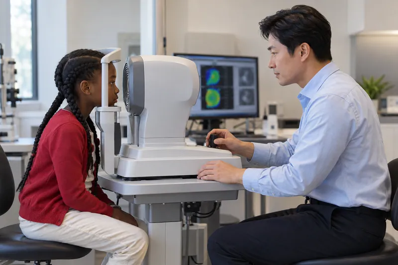
The fictional measurement scene illustrates acquisition, not a patient result. The display is not a diagnostic example. This article deliberately does not provide invented topographic patterns that could be mistaken for measured evidence.
Separate lens condition, care and ocular response
Before ordering a change, confirm which lenses the patient is wearing and inspect them as appropriate. Lens identity, condition, deposits, damage or an incorrect lens can change the investigation. Ask about handling and products without implying that an unfavorable result is automatically the family's fault.
Review the actual care process. Ask the patient or caregiver to describe what happens from removal through storage and the next application. A label that says “cleaned daily” may conceal an incompatible product or a missed disinfection step.
The CDC recommends keeping lenses and cases away from water, using recommended care products and maintaining an appropriate backup. These principles support infection prevention; the exact regimen still comes from the prescribed product and clinician. [4]
Document findings even when the family reports using the routine correctly. Ocular response and adverse effects require assessment on their own merits. A good care history is not proof that continued wear is appropriate.
VISUAL GUIDE 3 / 3
A useful finding leads to a specific next check
A map looks different
Quality, alignment, coverage, map type and scale
Question this can clarifyWhether the comparison is interpretableDaytime vision is different
Task, timing, refraction, ocular health and wear context
Question this can clarifyWhat performance problem needs investigationA lens seems to behave differently
Identity, condition, handling and prescribed design
Question this can clarifyWhether the actual lens and routine match the recordA proposed parameter change
Repeatable evidence and the exact fitting guide
Question this can clarifyWhy that change is appropriate and how it will be reviewedMake an adjustment with a question you can answer
When a modification is clinically appropriate, state what problem it is intended to address and how the response will be assessed. Avoid changing several factors without recording why, because the next review may become difficult to interpret.
Consider the manufacturer's guidance, repeatable measurements and the patient's actual visual experience together. The same descriptive map finding may have different implications across designs or contexts. A generic instruction such as “flatten this curve whenever this color appears” is not a safe substitute.
Give clear interim instructions. The family should know what correction to use, whether the existing lens may be worn, what to do if symptoms occur and when the next assessment will happen. Do not leave a lens order as the only recorded plan.
Check that relevant staff and the laboratory receive the correct version of the order. Distinguish an agreed change from a tentative suggestion discussed during troubleshooting. Closed-loop communication is especially useful when replacement lenses and follow-up are arranged by different people.
Keep refractive performance and progression monitoring distinct
A child may report clear daytime vision while axial measurements continue to change. Conversely, a visual-performance issue does not prove that a progression-control strategy has failed. The clinician needs to assess each question with appropriate methods.
Ortho-K alters corneal optics. Interpreting a routine refractive value as if the child were wearing only ordinary spectacles can be misleading. Record treatment status and the measurement context when discussing trends.
Use the monitoring guide to organize measurements and follow-up. Do not use unaided daytime acuity or an attractive map pattern as a stand-alone measure of progression control.
A decision to continue, adjust, pause, switch or refer should include a reason and a review plan. When the available data cannot answer the question, explain the uncertainty rather than converting it into a confident label of success or failure.
Teach the family what happens between visits
Families need instructions that can be used outside the consulting room. Confirm that the child and caregiver know the prescribed routine, relevant restrictions, symptoms requiring advice and the contact route when the practice is closed.
A review should check how care actually fits school, travel and sleep schedules. The response to a missed night or another disruption depends on the specific prescription. Provide individualized advice rather than relying on a generic online answer.
Preserve the distinction between a routine review and urgent assessment. A booked fitting follow-up is not a reason to delay care for a new painful or red eye. Record the safety advice given and confirm understanding.
The family should leave knowing what happens next, who will contact them about a replacement and what they should do if the plan becomes unclear. A technically appropriate lens order is only part of a complete clinical handoff.
Maintain the limits of a general troubleshooting guide
This page is a framework for organizing an investigation. It does not provide an individual lens design, diagnose a corneal condition or replace manufacturer training and professional judgment.
The American Academy of Orthokeratology and Myopia Control (AAOMC) offers practitioner education and professional resources. Use appropriate continuing training and design-specific support to maintain competence. Listing the Academy does not imply its endorsement of this article. [5]
For parent-facing safety questions, link families to the Ortho-K safety review alongside their actual instructions. Use the worksheet here to check the completeness of the clinical record and handoff.
Your fitting investigation
Discussion prompts only. Checks stay on this open page; nothing is submitted or saved. This sheet does not diagnose, choose treatment or determine urgency. Do not delay urgent care to complete it.
CHECK THE EVIDENCE
Sources & references
Research, professional guidance and patient resources. Checked October 22, 2026.
- 1Optimum Infinite Orthokeratology Lenses II: Professional Fitting and Information GuideManufacturer labeling in FDA PMA P240045 · 2026
- 2Optimum Infinite Orthokeratology Lenses II: Package Insert and Patient InstructionsManufacturer labeling in FDA PMA P240045 · 2026
- 3IMI: Myopia Management InstrumentationInternational Myopia Institute · IOVS · 2025
- 4Preventing Eye Infections When Wearing ContactsCenters for Disease Control and Prevention · 2025
- 5Practitioner Education and ResourcesAmerican Academy of Orthokeratology and Myopia Control (AAOMC)
General education. This article does not replace an eye examination, individual advice, or the prescribed care plan.